The problem: a handheld unit chosen on weight and price alone
A portable dental X-ray looks like a simple purchase: a light unit, a long battery life, no wall to drill. The backscatter shield turns out to be an optional extra, the exposure presets were written for film while the clinic uses a sensor, the round cone gives every patient a larger dose than a rectangular collimator would, and nobody has asked the regulator what it requires. This guide explains when a handheld, mobile-stand or wall-mounted unit fits, what the specifications mean for image and dose, why the receptor sets the exposure, how to protect operator and patient, and how the units we stock compare.
Understand: the unit, the numbers and the receptor
Handheld, mobile stand or wall-mounted
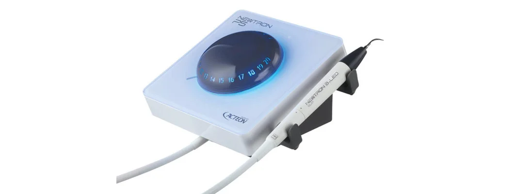
A handheld unit, sometimes sold as a handheld X-ray gun or portable X-ray camera, is a battery-powered tube head with a built-in cone and trigger, held by the operator for each exposure. It moves between surgeries and travels to domiciliary visits, special-care settings and outreach days without installation. A mobile-stand unit is a conventional mains-powered tube head on an articulated arm mounted on a wheeled stand: it rolls between rooms, the arm holds the tube head still, and the operator steps back to expose. The ADA and FDA recommendations state that dosimetry studies show hand-held devices present no greater radiation risk than standard units to the patient or the operator when used according to the manufacturer's instructions.1
Tube voltage, tube current and exposure time
Tube voltage, in kilovolts (kV), sets how penetrating the beam is and so the image contrast; the units in this guide work between 60 and 70 kV. Tube current, in milliamperes (mA), and exposure time together set how much radiation is produced. Battery handheld units run at low currents, such as the 2 mA of the Rolence portable unit, while the mains-powered Heliodent Plus runs at 7 mA. At a lower current the same receptor needs a longer exposure, which gives more time for movement, so compare exposure times, not only kV.
Focal spot
The focal spot is the area of the anode that emits the X-rays; the smaller it is, the less geometric unsharpness blurs enamel edges, root outlines and file tips. The Woodpecker Ai Ray and the Heliodent Plus both state 0.4 mm; some handheld tubes state larger figures.
Distance and collimation
The ADA and FDA recommendations note that a long source-to-skin distance of 40 cm, rather than a short distance of 20 cm, decreases patient exposure.1 Handheld cones are short, so ask for the stated source-to-skin distance. A round cone produces a circular field much larger than a sensor; a rectangular collimator trims the beam to roughly the receptor's shape. The same recommendations state that a rectangular collimator decreases the dose by up to fivefold compared with a circular one, and that equipment should provide rectangular collimation for periapical and bitewing radiographs.1 A systematic review of 13 studies found reductions of 40% to 92% compared with round collimation and rated the evidence as moderate.3 The trade-off is a greater risk of cone cuts when beam and receptor are misaligned, which is why holders that align the receptor with the collimated beam are recommended.1
The receptor sets the exposure
CMOS sensors are rigid, wired and show the image in seconds; size 1 suits anterior periapicals, children and narrow arches, and size 2 suits adult posterior periapicals and bitewings. Phosphor plates are thin, flexible and cable-free, and are read in a scanner after exposure. The ADA and FDA recommend the fastest receptor compatible with the diagnostic task: F-speed film reduces exposure by 20 to 50 percent compared with E-speed film, and digital imaging provides an opportunity to reduce the dose by a further 40 to 60 percent.1 Exposure settings therefore belong to the receptor, not to the X-ray unit. A preset written for film overexposes a sensor, and the wide exposure latitude of digital receptors means an overexposed image can still look acceptable on screen. Set the presets for your receptor by tooth region and patient size, and reset them whenever you change receptor.
Protecting the operator
The operator stands behind the backscatter shield, a leaded disc around the end of the cone. The ADA and FDA list the precautions the dosimetry depends on: holding the device at mid-torso height, orienting the shielding ring properly with respect to the operator, and keeping the cone as close to the patient's face as practical.1 In a laboratory study of one handheld unit used as its manufacturer specifies, no dose was detected on the handle or housing and no scattered radiation on the operator's side of a vertical plane through the focal spot; backscatter concentrated between patient and unit, within about 45 cm.4 Units differ, though. A phantom study of three handheld devices found operator doses that varied between models, recorded a finger dose of 16.76 mSv over two months for a dentist handling the sensor clinically, and recommended sensor holders, remote triggering, shielding discs and extended cones.5 Dental professionals should not hold the receptor during exposure; a family member or carer who must hold it or restrain the patient should wear appropriate shielding.1
Protecting the patient
Patient protection rests on justified radiographs, rectangular collimation, the fastest suitable receptor and a holder.1 Shielding advice has changed. The 2012 ADA and FDA recommendations advised protective thyroid collars whenever possible.1 The American Academy of Oral and Maxillofacial Radiology recommended ending routine thyroid and abdominal shielding in a 2023 position statement. In February 2024 an ADA expert panel report in JADA stated that thyroid and abdominal shielding during dental imaging is no longer recommended and should be discontinued as routine practice for all patients, pregnant patients included. The report adds that laws or regulations may still mandate certain equipment and that dentists should abide by the laws where they practise.2 In Saudi Arabia, radiation practices and facilities are regulated by the Nuclear and Radiological Regulatory Commission (NRRC), established in 2018 to protect people and the environment from radiation exposure.6 Confirm its requirements before the first exposure; clinics elsewhere in the GCC should ask their own regulator.
Battery, weight, stand and service
A handheld unit is held up for every exposure, so weight matters over a full-mouth series; the Ai Ray is listed at 2.4 kg. Exposures per charge vary widely between models: count your busiest day's radiographs, add a margin, and ask whether the clinic can replace the battery. A tripod with a remote exposure switch, where offered, holds the unit still and lets the operator step away. For any unit, ask who services it locally, how long the warranty runs, whether the backscatter shield is included, and whether a rectangular collimator is available. A handheld unit is touched with gloved hands at every exposure, so check that its grip and controls can take a barrier or be wiped, and buy sensor covers and autoclavable holders with it.
Options: the units and accessories we stock
| Unit | Type | Notable features (manufacturer) | Fits |
|---|
| Woodpecker Ai Ray | Handheld, battery | 0.4 mm focal spot, 2.8-inch touch screen, 2.4 kg, fast charging | Clinics sharing one unit between surgeries |
| Dexcowin DX3000 | Handheld, battery | Lead-infused backscatter shield, Toshiba tube; kV and cone length vary by version, so confirm them for the unit supplied | Domiciliary and outreach work |
| Rolence Portable X-Ray System | Handheld, battery | 60 kV, 2 mA, listed at over 450 exposures per charge; the maker lists the backscatter shield as optional | A second unit; confirm the shield is supplied |
| Dental Imaging Essentials Bundle | Handheld unit plus sensor | The Rolence portable unit with a Fussen F100 size 1 sensor | A clinic starting digital intraoral imaging |
| Dentsply Sirona Heliodent Plus, mobile stand | Mains tube head on a mobile stand | 60 to 70 kV, 7 mA, 0.4 mm focal spot; works with film, phosphor plates and sensors | A conventional unit moved between rooms |
| Item | Stocked examples | Role |
|---|
| Size 1 sensors | Xios AE, Fussen, NANOPIX1, Dental RVG Sensor H1 | Anterior periapicals, children, narrow arches |
| Size 2 sensors | Xios XG Select, Fussen, Dental RVG Sensor H2 | Adult posterior periapicals and bitewings |
| Holders | Rinn XCP-DS Fit Complete Kit (ORA + Endo), XCP Bitewing Kit #2 horizontal | Align receptor and beam; the XCP-DS Fit takes size 1 and 2 sensors |
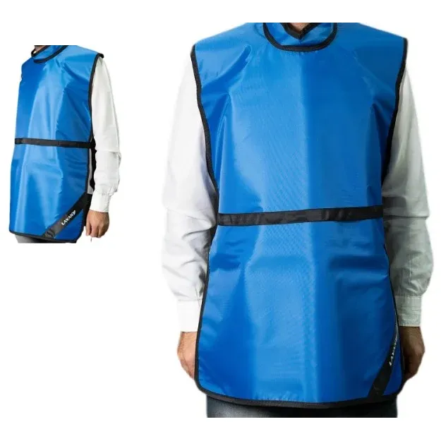
| Aprons and collars | Adult 0.35 mm Pb apron with collar, child aprons, 0.35 mm and 0.5 mm thyroid collars, apron hanger | Carers who must hold a receptor, and wherever the regulator requires shielding |
Buy the receptor, holders and covers with the unit.